Matching and Clustering
Introduction
The graph mining algorithms in the previous section tell us which PDBs a subgraph pattern is present in, but they do not tell us which specific residues are involved. Additionally, two subgraphs belonging to the same pattern can have different orientations or correspond to different residues in the crystal structure, and thus may not be related to one another in a meaningful way. In PyeMap, we cluster subgraphs based on their simiarlity in order to identify shared pathways/motifs.
Graph Matching
In order to identify the specific residues involved in subgraph patterns, we utitlize the NetworkX implementation of the VF2 algorithm for graph matching. We refer the reader to their documentation for more details, but for the sake of clarity, we re-iterate some key defintions.
Let G=(N,E) be a graph with a set of nodes N and set of edges E.
- If G’=(N’,E’) is a subgraph of G, then:
N’ is a subset of N,
E’ is a subset of E
- If G’=(N’,E’) is isomorphic to G, then:
there exists a one-to-one mapping between N and N’,
there exists a one-to-one mapping between E and E’
- For two graphs G(V,E) and H(V’,E’), G and H are subgraph isomorphic if there exists a G’(\(V_0,E_0`\)) such that:
G’ is a subgraph of G,
G’ is isomorphic to H
Note that in some sources, the term subgraph isomorphism is reserved for when G’ is a node- or edge-induced subgraph, and the term monomorphism is preferred for subgraphs which are not induced. The task of identifying all such G’ is known as the subgraph matching problem, and we refer to individual G’ as subgraph isomorphisms.
In PyeMap, for each PDB which supports the given subgraph pattern, we search for all subgraph isomorphisms within the protein graph. This gives us a set of protein subgraphs, which we can then cluster into groups based on similarity.
Each protein subgraph for a given subgraph pattern is assigned a unique ID with the format:
{PDB ID}-{Unique Index}
e.g. 1u3d-2.
Clustering
PyeMap currenly enables two types of clustering: structural similarity, and sequence simiarlity, which both use the same underlying algorithm below.
Algorithm
For a given subgraph pattern P, we have a set of protein subgraphs V which correspond to groups of specific residues in PDB structures which match pattern P. We then construct a supergraph G(V,E), where two protein subgraphs share an edge if and only if they are deemed sufficiently similar by the chosen metric. The resulting supergraph G will be composed of one or multiple connected components, and each connected component corresponds to a cluster of similar protein subgraphs.
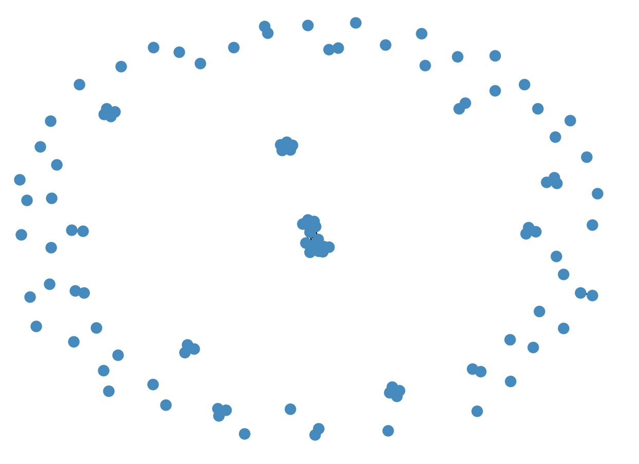
Fig. 3 Example supergraph G, where each node is a protein subgraph belonging to a subgraph pattern P. In this example, the supergraph G is composed of 66 connected components, and therefore, 66 clusters.
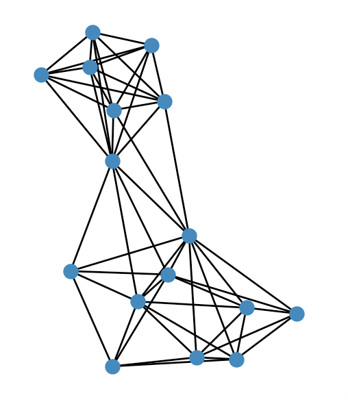
Fig. 4 The largest cluster in Fig. 3 zoomed in.
Structural Similarity
The structural similarity between two protein subgraphs in PyeMap is computed by superimposing the two sets of atoms
and computing the root mean squared distance (RMSD) using the Bio.PDB.Superimposer module in BioPython.
However, atoms can sometimes be missing from crystal structures, and we would also like some flexibility to allow for substitutions.
Starting from a one-to-one mapping between the residues, we make the following approximations to the true RMSD:
Only the alpha carbon (CA) is considered for standard amino acid residues. If it is not present in the crystal structure, \(\infty\) is returned.
For non-standard amino acids, we use the first atom type both residues have in common. If no shared atom type is found, \(\infty\) is returned.
An edge is drawn between two protein subgraphs in the supergraph if RMSD \(\leq\) 0.5 Å.
Sequence Similarity
Sequence simiarlity in PyeMap relies on a multiple sequence alignment, which will automatically be performed by the MUSCLE package [Edgar2004] if it is installed on your machine. Starting from a one-to-one mapping between the residues, the sequence similarity between two protein subgraphs is simply defined as the sum of the differences in the residue numbers with respect to the multiple sequence alignment. For instance, TRP50 in one PDB and TRP200 in another PDB could have a difference of 0 if they are aligned by the multiple sequence alignment. One important caveat is that non-protein residues are not considered in sequence similarity, only standard amino acid residues.
An edge is drawn between two protein subgraphs in the supergraph if \(D \leq N\), where D is the sum of the differences in the residue numbers with respect to the multiple sequence alignment, and \(N`\) is the total number of nodes comprising the subgraph pattern, which allows for slight misalignments.
Note:
If MUSCLE is not installed, the original residue numbers will be used, which is unlikely to lead to a meaningful clustering.
Source
Compute the supergraphs and find the connected components |
|
Computes RMSD between two protein subgraphs |
|
Computes sequence distance between two protein subgraphs |